Raman spectroscopy is a versatile method that is mainly used for particle analysis at our Dresden business location. Particles of up to one diameter of 1 µm (both inorganic and organic) that exhibit Raman activity can be identified here.
In addition, so-called high-resolution line scans can be used to scan surfaces and to view two-dimensional contamination using optical and spectroscopic methods.

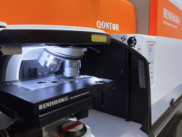
Monochromatic light from a laser that has a wavelength of either 532 or 785 nm is used to analyze a sample. The light is directed through an objective onto a sample and scattered after interaction with the sample. A shift of the initial wavelength then takes place (so-called Raman shift). After detection, these shifts are displayed in a spectrum and are characteristic for molecular groups within the sample. A change in polarizability within the molecules must be possible (RAMAN-active) in order for this method to be applied. Samples that fluoresce strongly due to their molecular structure or coloration cannot be identified in some cases because the detector is unable to distinguish between Raman and fluorescence radiation.
Raman spectroscopy is often used in addition to FT-IR spectroscopy or REM-EDX analysis for more accurate substance group identification.